

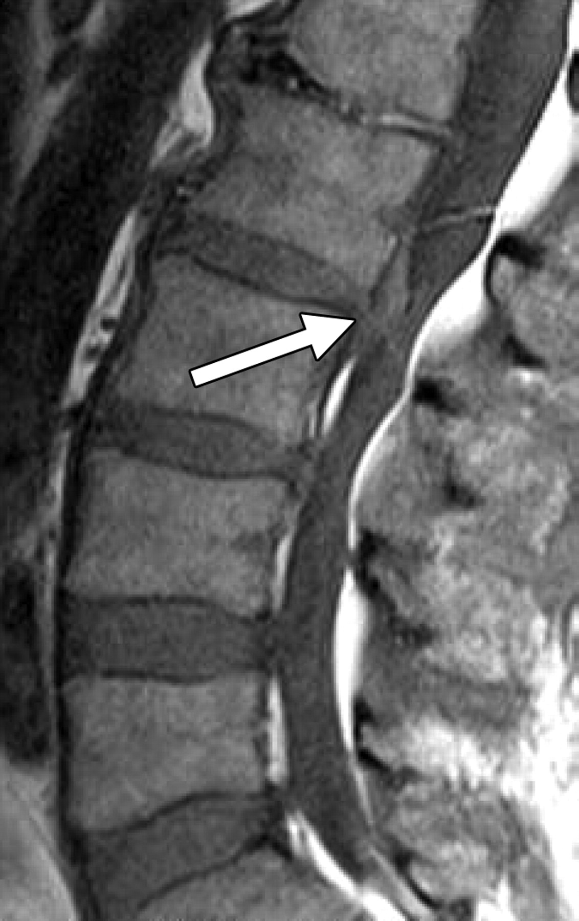
What can an MRI scan of the spine diagnose?
An MRI scan of the spine is a detailed scan which shows the health of spinal bones, discs, nerves and ligaments. Unlike a CT scan or x-ray it is a non-radiation scan making it low risk scan.
As doctors we often request MRI scans when we suspect that problems with the spine may be causing our patients neck pain, back pain, stiffness or mobility problems.
60-80% of the population experience back pain at some point in their lives and the vast majority of these people manage their pain without any input from their doctor or physiotherapist. However in some cases an MRI scan is a vital test which diagnoses the exact cause of a patient's pain symptom. The results of an MRI scan are even more vital when planning rehabilitation such as physiotherapy or even surgery for spinal problems.
Which parts of the spine can be scanned with MRI scans?
An MRI scan provides detailed images of the:
-
Cervical spine (neck)
-
Thoracic spine (mid-back)
-
Lumbar spine (lower back)
-
Intervertebral discs
-
Spinal cord
-
Nerve roots
-
Ligaments and soft tissues
MRI is particularly effective at detecting problems involving nerves, discs, and soft tissues that may not be visible on other tests such as x-rays.
Which symptoms may warrant an MRI scan?
You may wish to have an MRI scan for the following symptoms;
Back Pain
If back pain continues despite treatment or is affecting daily activities, an MRI can help identify the underlying cause.
Neck Pain
MRI scans can reveal disc problems, nerve compression, inflammation, and other conditions affecting the cervical spine.
Sciatica
Sciatica occurs when irritation or compression of a nerve causes pain, numbness, or tingling that travels down the leg. MRI is often used to identify the source of nerve compression.
Slipped disc
A herniated disc can press on nearby nerves, causing pain, weakness, or altered sensation. MRI provides detailed images of spinal discs and surrounding structures.
Spinal stenosis
This condition involves narrowing of the spinal canal, which can place pressure on the spinal cord or nerves.
What can I expect during the MRI scan?
The MRI scanner can be considered a "huge magnet" therefore you should expect to remove any metal jewellary including piercings before the scan.
During the scan;
-
You will lie on a scanning table.
-
The table moves into the MRI scanner.
-
The scanner takes a series of images.
-
You must remain as still as possible.
-
You will hear loud tapping or knocking sounds during image acquisition.
Many imaging centres provide ear protection to improve comfort during the procedure.
For further information;
Visit FAQs page for more information on how to book a scan through Skannr
Visit the MRI scan page to choose a location near you and make an MRI scan booking.